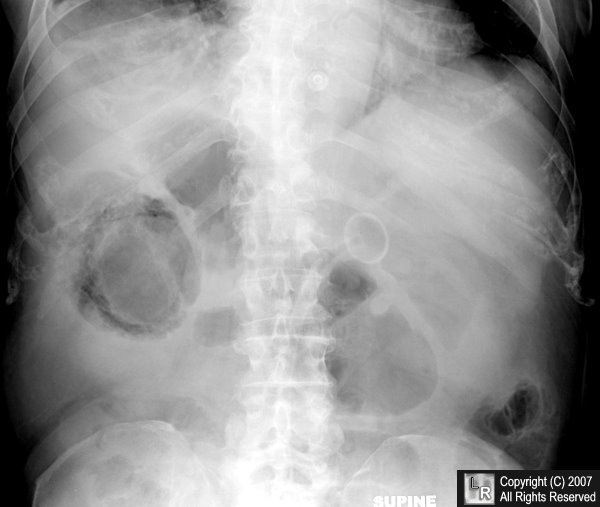
黄色肉芽腫性胆嚢炎
INTRODUCTION — Xanthogranulomatous cholecystitis is a rare inflammatory disease of the gallbladder characterized by a focal or diffuse destructive inflammatory process, with accumulation of lipid laden macrophages, fibrous tissue, and acute and chronic inflammatory cells [1] . In 1970 it was first known by the descriptive term fibroxanthogranulomatous cholecystitis [2] but in 1981 the name xanthogranulomatous cholecystitis was proposed in a review of 40 cases from the Armed Forces Institute of Pathology [3] . Its importance lies in the fact that it is a benign condition that may be confused with carcinoma of the gallbladder, which is associated with a poor prognosis.
Xanthogranulomatous cholecystitis was initially described as a variant of chronic cholecystitis. However, while the latter is usually regarded as a benign condition with questionable clinical significance, xanthogranulomatous cholecystitis is an active and destructive process that can lead to significant morbidity as the inflammatory process usually extends into the gallbladder wall and adjacent structures. Thus, it should be considered a distinct clinical entity.
EPIDEMIOLOGY — The prevalence of xanthogranulomatous cholecystitis among patients with symptomatic gallbladder disease ranges from 0.7 percent in the United States to up to 10 percent in India and Japan [4-8] . The mean age at presentation varies in different studies from 44 to 63 years [4,9] . A study from India reported a 1:9 male to female ratio [4] while in other reports the male to female ratio ranged from 2:1 to 1:2 [3,7,9] .
PATHOGENESIS — The pathogenesis of xanthogranulomatous cholecystitis is thought to be related to extravasation of bile into the gallbladder wall from rupture of Rokitansky-Aschoff sinuses or by mucosal ulceration [3,10] . This event incites an inflammatory reaction in the interstitial tissue whereby fibroblasts and macrophages phagocytose the biliary lipids in bile, such as cholesterol and phospholipids leading to the formation of xanthoma cells.
Gallstones may have an important role in the pathogenesis, since they appear to be present in all patients [4,9,11] . It has been suggested that xanthogranulomatous cholecystitis is analogous to xanthogranulomatous pyelonephritis, which results from obstruction and stasis due to renal calculi [12] .